


Age-related Eyebrow and Eye Ptosis
Aesthetic and Plastic Surgery Specialist Op. Dr. Ufuk Askeroğlu explained for us what is curious about eyebrow and eye ptosis, which is the problem of many people today.
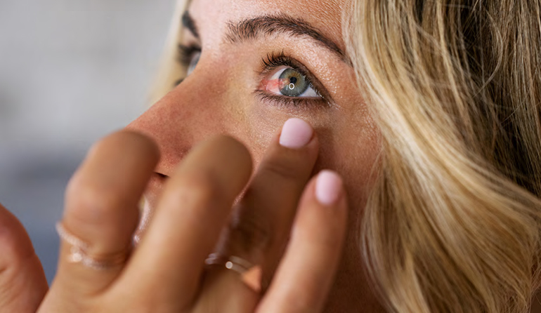
Eyebrow and eye ptosis has become a problem experienced by many people today. This drooping can be seen in one or both eyes. In some cases, a slight sagging occurs, and in some cases, the sagging of the eyebrows or eyelids can fall down to the level of the pupils. In these and similar cases, vision is restricted and patients may experience vision loss. Eyebrow tissue is very soft and can weaken. Weak eyebrows can cause a tired appearance. In addition to appearance, it can also cause visual discomfort.
Various brow lift methods can make you look younger and more energetic. Eyebrows have a great effect on facial expressions. Asymmetrical and drooping eyebrows can reduce expression and cause self-confidence problems.
There may be more than one cause of droopy eyelids. Eyelid drooping occurs with the weakening of the eyelid muscles. As we age, the muscles that lift and hold the eyelids open weaken. Weak muscles become heavier and come under the influence of gravity. In some cases, the disorder can be congenital. During development in the womb, the muscles that open the eyelids may be underdeveloped.
How to treat droopy eyelids and the accompanying problems?
In the Bella Eyes procedure, the eyebrows are lifted by pulling them up and the eyes appear more slanted than they are. This procedure; It is preferred by people who have started to fall from the edges of the eyes, crow’s feet and eyelids. Thanks to the application, people can get rid of these problems and achieve the vibrant and youthful appearance they want. Trinity Lift surgery is also frequently performed in combination with Bella Eyes. Since both are performed through the same incision, both mid-face and brow lift are achieved at the same time. Since it is a surgical procedure, it must be performed by an expert plastic surgeon. The biggest advantage is that it leaves no scars and the incision remains completely in the scalp. The operation can be performed in the hospital overnight and the patient can return to normal life within 7 days on average.
Pay attention to these after the procedure!
Some conditions must be taken into consideration for a fast and completely scarless recovery after Bella Eyes application. Since this procedure is not a very long and difficult surgical intervention, patients are not expected to experience serious complications, but faster recovery can be achieved by paying attention to certain points.
- It is recommended not to lie face down for about a week.
- Doctor’s recommendations and warnings must be followed.
- Ice should continue to be applied on the first day. It helps to remove swelling and bruises in the area.
- Swelling on the face after surgery will naturally disappear in about 1 week.
- The surgical scars formed during the surgery should be protected from sunlight and sunbathing for 2-3 months.
September 2024